
Correlation study of quantitative and qualitative analysis results in PET/CT imaging of temporal lobe epilepsy patients in Iran
Given the 50% sensitivity of PET images for diagnosing temporal lobe epilepsy, the results obtained from quantitative analyses are effective in confirming and helping to diagnose the area of involvement.
Dr. Mohammad Reza Ay
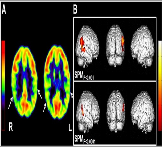
Given the 50% sensitivity of PET images for diagnosing temporal lobe epilepsy, the results obtained from quantitative analyses are effective in confirming and helping to diagnose the area of involvement. Given that diagnosing the area of involvement may ultimately lead to the diagnosis of the epileptic focus and surgery for that area, ensuring the accuracy of the visual diagnosis is very crucial. Currently, one of the best methods to help visually diagnose temporal lobe epilepsy is the numerical analysis of different brain areas. With quantitative analysis, doctors can evaluate the results and reach a more accurate conclusion about the focus of involvement. The purpose of this study is to examine and compare the results of quantitative analysis of brain PET images obtained using special brain analysis software with the PET reports kept by doctors.

comment